This manual provides a structured learning experience for anatomy and physiology, offering hands-on activities, safety guidelines, and exercises tailored for healthcare students. Designed to accompany any A&P textbook, it is available in three versions (Main, Cat, and Fetal Pig) to suit diverse learning needs.
1.1 Purpose and Scope of the Manual
This manual serves as a comprehensive guide for anatomy and physiology labs, designed to help students explore human structure and function through hands-on activities. It aligns with a two-semester course, covering all body systems with exercises, dissections, and physiological experiments. The scope includes supporting healthcare-focused learning and integrating with various textbooks for a tailored educational experience.
1.2 Versions of the Manual (Main, Cat, Fetal Pig)
The manual is available in three versions to accommodate diverse learning needs: the Main version for general use, the Cat version with nine cat dissection exercises, and the Fetal Pig version for comparative anatomy. Each version provides tailored activities, ensuring students gain practical insights into human and comparative anatomy through structured exercises and dissections.
Laboratory Safety and Equipment
This section emphasizes the importance of safety protocols and proper equipment handling, providing foundational knowledge and practical guidance for a safe and effective learning environment.
2.1 Essential Safety Protocols
Adhering to safety protocols is crucial in anatomy and physiology labs. Students must wear protective gear, handle chemicals and instruments safely, and follow proper disposal procedures. Emergency exits and equipment should be easily accessible. Clear communication and adherence to instructor guidelines ensure a secure environment, preventing accidents and fostering a focused learning atmosphere.
2.2 Common Laboratory Equipment and Tools
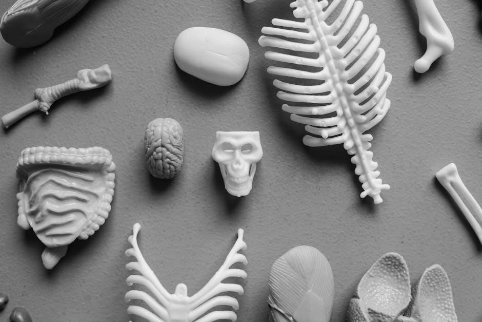
Essential lab equipment includes microscopes for tissue examination, dissection tools like scalpels and forceps, and measurement tools such as calipers and thermometers. Anatomical models and skeletal specimens aid in understanding structures. Laboratory glassware, including beakers and test tubes, supports physiological experiments. The manual provides detailed guidance on the safe and effective use of these tools to enhance hands-on learning experiences.
This section introduces foundational concepts of the human body, including anatomical terminology, body systems, and the relationship between structure and physiological function.
3.1 Anatomical Terminology
Anatomical terminology provides a standardized language for describing body structures and their locations. Terms like “ventral,” “dorsal,” and “proximal” help identify positions and orientations. Understanding this vocabulary is essential for accurate communication in anatomy and physiology, enabling precise identification of organs, tissues, and systems. The manual includes visual aids and exercises to master these terms, ensuring a strong foundation for further study and practical applications in healthcare fields.
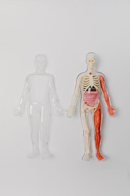
3.2 Body Systems Overview
The section provides a comprehensive overview of the body’s major systems, such as skeletal, muscular, and nervous. It includes detailed descriptions of system functions, interactions, and homeostatic mechanisms. Exercises, diagrams, and clinical correlations enhance understanding. The manual’s structured approach helps students connect anatomical structure with physiological function, essential for healthcare applications. This foundation supports advanced topics and ensures a cohesive learning experience.
Tissues and Histology
This section introduces the four primary tissue types—epithelial, connective, muscle, and nervous—and explores histological techniques for preparing and examining tissue samples under a microscope.
4.1 Types of Tissues
The four primary types of tissues are epithelial, connective, muscle, and nervous. Epithelial tissues form linings and glands, while connective tissues provide support and structure. Muscle tissues enable movement, and nervous tissues facilitate communication. Each tissue type has distinct functions and structures, contributing to the body’s overall functionality. The manual includes histological images and exercises for identifying and understanding these tissues in detail.
4.2 Histological Slide Preparation and Microscopy
Histological slide preparation involves sectioning, staining, and mounting tissues for microscopic examination. Staining enhances contrast, revealing cellular structures. Proper microscopy techniques, such as focusing and adjusting magnification, are essential for accurate observations. The manual provides step-by-step guidance and tips for identifying tissue structures, ensuring a clear understanding of histological features. This process is fundamental for studying tissue composition and function.
Integumentary System
The integumentary system focuses on the structure and function of skin, exploring its layers, accessory organs, and protective roles. Laboratory exercises include histological examinations and sensory receptor studies.
5.1 Structure and Function of the Skin
The skin, the body’s largest organ, consists of the epidermis, dermis, and hypodermis. It protects against external damage, regulates temperature, and aids in sensation. Laboratory exercises focus on histological examinations of skin layers, identification of accessory organs like hair follicles and sweat glands, and understanding the skin’s role in maintaining homeostasis and immune defense.
These activities enhance comprehension of its complex functions.
5.2 Laboratory Exercises on Skin and Accessory Organs
Exercises involve microscopic examination of skin histology, exploring epidermal layers, dermal components, and hypodermal tissues. Students identify accessory organs like hair follicles, sebaceous glands, and sweat glands. Magnification techniques reveal structural details, while dissection activities provide hands-on experience with skin thickness and sensory receptor locations, enhancing understanding of its protective and sensory roles.
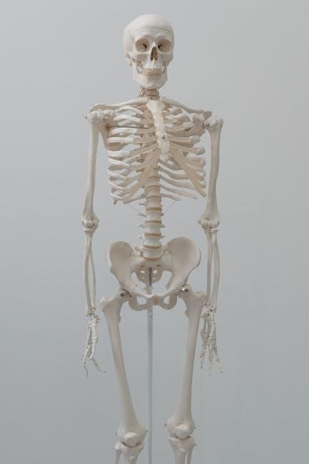
Skeletal System
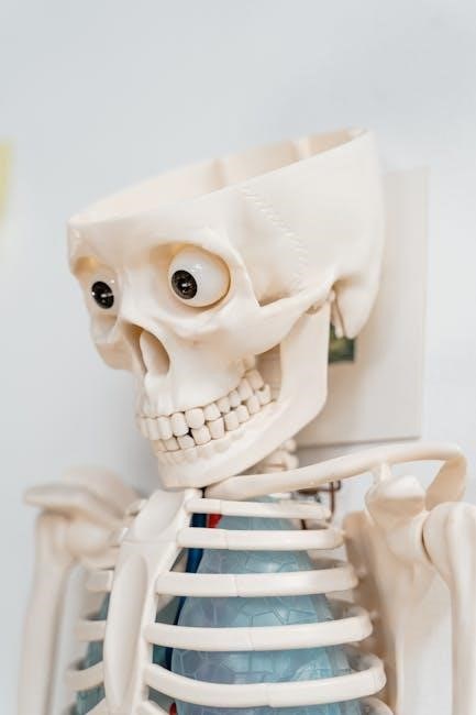
This section explores the structure and function of bones and joints, guiding students through exercises on skeletal anatomy, including detailed studies of the skull and vertebral column.
This section introduces the fundamentals of the skeletal system, focusing on bone classification, structure, and function. It explores types of joints, their movements, and stability mechanisms. Exercises include identifying bone features and understanding joint anatomy through dissection and microscopy. The manual provides detailed illustrations and hands-on activities to enhance comprehension of skeletal components and their role in movement and support.
6.2 Detailed Study of the Skull and Vertebral Column
This section focuses on the intricate anatomy of the skull and vertebral column, emphasizing their structural and functional significance. Exercises include identifying cranial and facial bones, understanding vertebral types, and exploring spinal curvature. Detailed illustrations and dissection guides facilitate hands-on learning, while study guides and microscopy enhance comprehension of these critical skeletal components.
Muscular System
This section explores the structure and function of muscles, including skeletal, smooth, and cardiac types. Lab exercises focus on muscle identification, dissection, and understanding their roles in movement.
7.1 Types of Muscles and Their Functions
The muscular system comprises three types of muscles: skeletal, smooth, and cardiac. Skeletal muscles attach to bones, enabling voluntary movements and supporting posture. Smooth muscles, found in internal organs, facilitate involuntary actions like digestion. Cardiac muscle, exclusive to the heart, ensures rhythmic contractions for blood circulation. Each type has distinct structures and functions, crucial for overall bodily movement and physiological processes.
7.2 Dissection and Identification of Major Muscle Groups
Dissection and identification of major muscle groups provide hands-on experience in understanding their structure and function. This exercise involves carefully examining and labeling muscles in specimens, focusing on groups like flexors, extensors, and adductors in the upper and lower limbs, as well as abdominal muscles. Students learn to use scalpels and forceps to explore muscle attachments, fiber directions, and how muscles contribute to movement and posture. This practical approach reinforces anatomical knowledge and its physiological significance.
Nervous System
This section explores the structure and function of the nervous system, focusing on neural tissues, reflexes, and laboratory exercises to understand its role in controlling bodily functions.
8.1 Basic Structure and Function
The nervous system consists of neurons and glial cells, functioning to transmit and process information. It is divided into the central (brain and spinal cord) and peripheral systems, enabling communication, control, and coordination of bodily functions through electrical and chemical signals, forming the basis of movement, sensation, and cognitive processes.
8.2 Laboratory Exercises on Neural Tissues and Reflexes
Lab exercises explore neural structures through dissection and histological slides, examining brain and spinal cord anatomy. Students study reflex arcs, testing responses to stimuli, and analyze nerve impulse conduction. These hands-on activities integrate theoretical knowledge with practical skills, enhancing understanding of neural function and its role in controlling body systems.
Clinical Applications and Case Studies
This section bridges anatomy and physiology with real-world healthcare scenarios, offering case studies that illustrate physiological disorders and their clinical relevance, enhancing practical understanding for healthcare students.
9.1 Linking Anatomy and Physiology to Real-World Scenarios
This section connects anatomical structures and physiological processes to clinical scenarios, enabling students to apply theoretical knowledge in practical healthcare contexts. Through case studies and examples, learners develop critical thinking skills to diagnose and understand common disorders, fostering a deeper appreciation of how body systems function in health and disease.
9.2 Common Disorders and Their Physiological Basis
This section explores the physiological mechanisms behind frequent health disorders, such as diabetes, hypertension, and respiratory conditions. It examines how disruptions in normal bodily functions contribute to disease development, emphasizing the interplay between body systems. Through detailed explanations and case studies, students gain insights into the clinical relevance of anatomical and physiological principles, enhancing their understanding of human health and disease.
Study Resources and Tools
This section provides supplementary materials, including review sheets and interactive activities, to enhance learning. Online platforms like Mastering A&P offer additional practice and visualization tools for complex topics.
10.1 Recommended Supplementary Materials
Supplementary materials include textbooks like Marieb/Hoehns Human Anatomy & Physiology, online platforms such as Mastering A&P, and interactive tools like eLabs. These resources provide additional practice, quizzes, and visual aids to reinforce learning. They also offer detailed dissection guides, histology slides, and clinical case studies to enhance understanding and practical skills in anatomy and physiology.
10.2 Online Platforms for Interactive Learning
Online platforms like Mastering A&P and eLabs offer interactive simulations, quizzes, and virtual dissection tools. These resources provide 3D models, video tutorials, and adaptive learning exercises to enhance engagement. They cater to diverse learning styles and supplement lab manual content, allowing students to explore complex anatomical structures and physiological processes in a dynamic, self-paced environment.
Laboratory Report Writing and Presentation
The manual aids in documenting experiments effectively, providing clear instructions, safety tips, and tear-out worksheets. Additional interactive resources, like videos and tutorials, enhance understanding and presentation skills.
11.1 Guidelines for Documenting Lab Experiments
The manual provides clear guidelines for documenting lab experiments, emphasizing organized and detailed reporting. It includes tear-out worksheets, safety tips, and step-by-step instructions to ensure accurate and professional documentation. Students are encouraged to record observations, data, and conclusions effectively, fostering a structured approach to lab reporting and scientific communication. This section helps in maintaining clarity and precision in experimental documentation, essential for healthcare-related studies and future reference.
11.2 Tips for Effective Scientific Communication
Effective scientific communication involves using clear and concise language, organizing thoughts logically, and incorporating visual aids to enhance understanding. Practice active listening and provide constructive feedback in group settings. Utilize resources from the lab manual, such as tear-out worksheets, to prepare well-structured presentations and reports. Regularly review and refine your communication skills to ensure clarity and professionalism in all interactions.
Instructor and Student Support
This section provides instructors with customizable content options and additional resources, while students benefit from interactive online platforms and supplementary study materials, enhancing both learning and teaching experiences.
12.1 Customizing the Lab Manual for Specific Courses
The manual allows instructors to tailor content to their course needs, offering flexibility in lab exercises and dissection activities. It supports customization through digital options and alignment with specific course goals, ensuring a personalized learning experience for students while maintaining core anatomical and physiological concepts. This adaptability makes it suitable for various healthcare and science programs, including nursing and allied health fields.
12.2 Additional Resources for Instructors
The manual offers supplementary materials for instructors, including digital tools, customizable exercises, and access to online platforms. These resources support lesson planning, student engagement, and assessment. Instructors can utilize videos, tutorials, and interactive activities to enhance teaching. Additional support materials, such as answer keys and lab setup guides, are also provided to facilitate effective instruction and management of anatomy and physiology courses.
The manual concludes by summarizing key concepts and emphasizing the importance of hands-on learning. Future updates will integrate emerging technologies and innovative teaching methods to enhance anatomy and physiology education.
13.1 Summary of Key Concepts
This manual provides a comprehensive overview of anatomy and physiology through structured exercises and dissections. It covers tissues, body systems, and laboratory techniques, emphasizing hands-on learning and critical thinking. The integration of histology, microscopy, and clinical applications prepares students for healthcare careers. Supplementary resources and online tools enhance understanding, reinforcing the connection between anatomical structure and physiological function.
13.2 Advances in Anatomy and Physiology Education
Modern advancements integrate digital tools, interactive simulations, and virtual dissections, enhancing traditional lab experiences. Online platforms and AI-driven resources now offer personalized learning paths. The rise of 3D modeling and augmented reality provides immersive explorations of anatomical structures. These innovations ensure anatomy and physiology education remains dynamic, accessible, and tailored to diverse learning styles, preparing students for future challenges in healthcare.